Offshore Physiotherapy Torquay

Torquay Clinic

144 Surfcoast Hwy, Torquay

Grovedale Clinic

222 Torquay Road, Grovedale

Women’s Health Physio

Women’s Health Physio

Offshore Physio Torquay – March 2020

Did you know that Offshore Physio has a physiotherapist specialising in Pelvic Floor rehab and pre and postnatal conditions?

Courtney Weybury has over 10 years of physio experience working in private practice and has Postgraduate qualifications in Pelvic Floor Rehabilitation & Continence and Exercise for Women.

Incontinence affects the lives of almost 5 million Australians and recent studies show that up to 70% of people living with the condition fail to seek help or advice. After childbirth 1 in 3 women have urinary incontinence and 1 in 2 have some degree of pelvic organ prolapse. Urinary incontinence can be stress, frequency, urge or mixed. The prevalence of urinary incontinence increases with age and is most commonly caused by a weakened pelvic floor due to pregnancy, menopause, obesity and other conditions such as constipation and chronic cough. Incontinence is also common in men post prostate surgery.

The pelvic floor muscles are important to support your pelvic organs (bladder, uterus and bowel) and to maintain bladder and bowel continence. The pelvic floor muscles are also important for sexual function and work in conjunction with the abdominal muscles to support and stabalise the spine.

When returning to sport and exercise post baby, it is important to consider what type of exercise you do initially and how quickly you go back to high impact sport and exercise. It is important to make sure your pelvic floor and stomach muscles are strong enough to support your spine and pelvis and to be able sustain the stresses from exercise by supporting the pelvic floor organs.

Incontinence can be treated and managed with pelvic floor muscle training and lifestyle modifications. Women’s health physiotherapy is a highly evidence based, specialised field of physiotherapy that can help treat incontinence, pregnancy related conditions, pelvic pain and dysfunction and post prostate surgery.

If you have any questions or would like more information regarding incontinence, pre and post natal conditions, pelvic pain or dysfunction or returning to sport, call us on 5261 5246 to book an appointment with Courtney.

Medical Imaging and back pain

Medical Imaging and back pain – what does the evidence tell us?

Offshore Physio Torquay – May 2019

An issue that often presents in the clinic is the decision on whether a patient requires a scan for low back pain (LBP). Patients often already arrive with scans on their first visit either from referral from another health care practitioner or from previous onsets. Some people are very fearful of results of scans, especially of the dreaded “disc Bulge”. This fear is perpetuated via google searches, discussions with friends and family who have had back pain and sometimes even by other health care professionals. However, the evidence tells us that what we see on scans has poor correlation to the amount of pain we experience, and therefore we shouldn’t fear a disc bulge. In fact, its more common than you think in people who do not have any pain. I thought it would therefore be appropriate to discuss the evidence behind the value of imaging for LBP.

What is LBP and how common is it

LBP is pain between the lower rib margins and the buttock crease and can occur with or without neurological referral into the lower limb. LBP is very common, affecting up to 80% of the population at some point throughout their lifetime. Therefore, it is more common than uncommon to have LBP at some point in your life. 90 to 92% of most LBP cannot be attributed to a specific structure and garners the term non- specific low back pain. The remaining presentations can be attributed to vertebral compression fractures (4%) or inflammatory spine diseases (<5%) (Wáng et.al, 2018). Less than 1% can then be accounted for by more serious underlying conditions such as infection (0.01%), Cauda equina syndrome (0.04%) and metastatic cancer (0.7%) (Wáng et.al, 2018). Signs of these more sinister presentations in the 1% of the population of LBP can be screened and monitored via physiotherapists and doctors through physical and subjective exam. It is this 1% where early imaging is vital (Wáng et.al, 2018).

How prevalent are change such as disc degeneration, disc bulges and facet arthritis

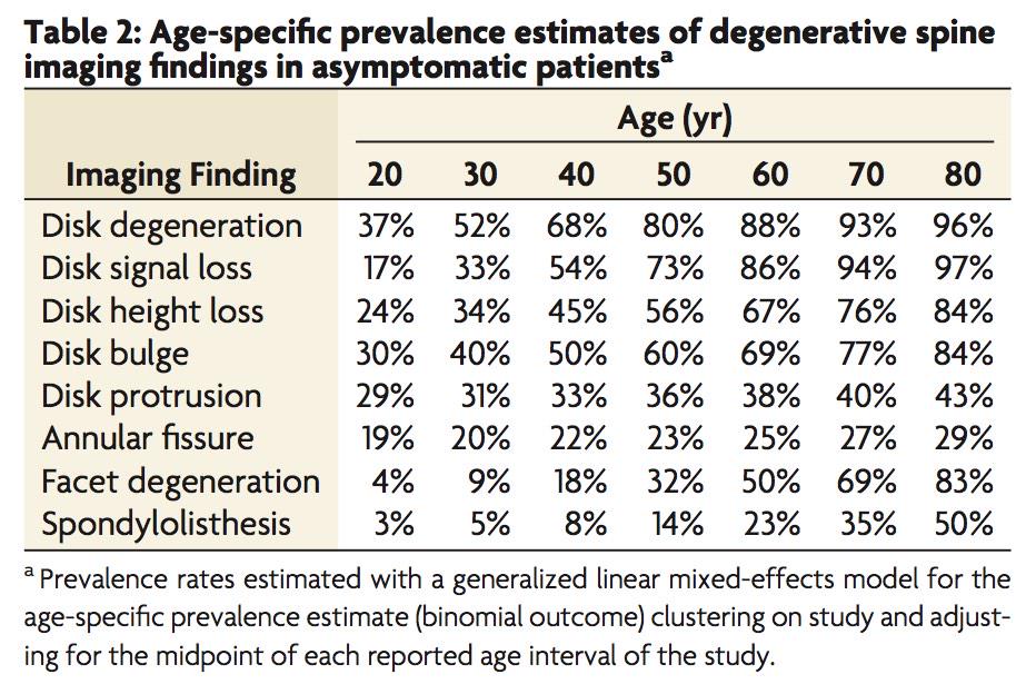
Studies have shown a poor correlation between structural changes we see on a scan and the level of pain a person will experience. Major evidence from this has come from studies that have scanned the backs of people whom do not have low back pain (asymptomatic). A Systematic review by Brinjkji et.al 2015 summarised the findings of all these studies. Some Major findings where that disc bulges are present in 60% of 50-year old’s (Brinjkji et.al 2015). Remember these are people who have no low back pain but almost two thirds will have a disc bulge (See Table 2 below for full results of the systematic review). Let’s put this into perspective. The chances are that based on this data most of us are walking around with some changes in our spine, but don’t have any pain. Therefore, when we do have an onset (which is highly likely as 80% of the adult population will experience LBP at some point in their lifetime), if we scan the back we may then blame the pain on a change in the spine that was highly likely to be their prior to the onset of pain, and is likely poorly correlated with the pain experience. The major conclusions of these studies are that changes on MRI for the non-specific LBP group cannot be interpreted as a direct cause for pain. Its not to say they do not contribute, its more that they may become sensitized rather than a direct cause to pain based on the degree of change.

(Brinjikji, et al., 2015, p.813)

Early scanning does not provide greater outcomes for LBP

Many people present expecting that they need a scan in order to determine appropriate treatment and enhance the chance to recover. The evidence however tells us that most of the time this is not the case (Darlow, 2017). In fact, a number of studies have shown worse outcomes in terms of pain and disability for patients who have had early imaging after LBP onset compared to those who didn’t or where blinded to the results of their scan until after the trial (Wáng et.al, 2018). Rationale for worse outcomes in these patients whom underwent early scanning may be that it produced unintended harms of diagnostic labelling, causing patients to embody these labels. These labels or clinically irrelevant findings can produce worse outcomes as it can cause the person with LBP to worry more, focus excessively on their pain or avoid exercise or return to work due to the fear of doing structural damage to their back (Wáng et.al, 2018). On top of this early scanning within a month of the onset of LBP was associated with 8 times greater risk for surgery and 5 times the amount of medical costs compared to those whom don’t undergo early imaging (Wáng et.al, 2018).

What are the guidelines for scanning low back pain

The guidelines for when to image the spine include:

  • LBP and radiculopathy (neurological symptoms) for greater than 6 weeks, after failure of conservative care (Physiotherapy)
  • Low back pain with severe, progressive neurological deficits (Loss of strength, power or reflexes)
  • Signs of a serious or specific underlying condition (cancer, infection or cauda equina) but remember this is less than 1% of LBP presentations, however we should always be vigilant in assessing for these presentations.

Despite this there has been a significant increase in MRI scanning over the last 15 years despite guidelines suggesting routine scanning is unhelpful.

Summary

There is strong evidence that routine imaging for LBP in unnecessary and can have more negative than positive effects on outcomes in terms of pain and associated disability. This is because many changes detected are normal and poorly correlated with pain. Early imaging in these cases therefore exposes patients to an increase risk of more invasive medical interventions and can lead to worry, focus on pain, protective movement and withdrawal from exercise, social activities and work. All of which can increase pain. Essentially for the 90 to 92% of patients the findings on scanning will not change how a physiotherapist helps you to manage your pain. A major caveat however is that early scanning is vital in the 1% of the population whom may present with infection, metastatic cancer or cauda equina. Ongoing screening through history taking and physical exam for these presentations should occur by a health professional.

Ryan Dahlhaus has been a physiotherapist since graduating in 2012 from Latrobe University. He has worked at Offshore Physio since 2015 and is currently completing his Masters of Science in Medicine in Pain Management at the University of Sydney. He works with a lot of people with chronic pain to get them back and moving. If you want to discuss a chronic pain condition, Ryan works at both the Torquay and Grovedale clinic.

References:

The information presented in this summary is from three main academic articles links to the full text are provided as all are open access papers:

Brinjiki W, Leutmer PH, Comstock B, et.al, (2015). Systematic literature review of imaging features of spinal degeneration in asymptomatic populations. AJNR Am J Neuroradiol 2015; 36 (4):811-6 https://www.ncbi.nlm.nih.gov/pmc/articles/PMC4464797/pdf/nihms-696022.pdf

Darlow, B., Forster, B., O’Sullivan, K., & O’Sullivan, P. (2017). It is time to stop causing harm with inappropriate imaging for low back pain. British Journal of Sports Medicine, 51(5), 414–415. https://bjsm.bmj.com/content/51/5/414.long

Wáng, Y., Wu, A., Ruiz Santiago, F., & Nogueira-Barbosa, M. (2018). Informed appropriate imaging for low back pain management: A narrative review. Journal of Orthopaedic Translation, 15, 21–34. https://doi.org/10.1016/j.jot.2018.07.009

Tips for Injury Free Running

Tips for Injury Free Running

Written By Ellie O’Kane Physiotherapist-  Offshore Physio Torquay – January 2019

So, first of all, I want to encourage everybody to run! There’s a misconception that some peoples “bodies aren’t made for running”, “I’m too old to run”, or “running is bad for your knees”. 

Well I’d like to quash those thoughts and replace them with ways to help you to run safely and with minimal injury risk. These tips are relevant to those who have never run in their lives, and those who run every day. If taking up running is done in a gradual and consistent manner the benefits are endless on both physical health (no it is not bad for your joints) and mental health (endorphins, stress relief, sense of achievement +++). It’s time efficient and it’s free (aside from the cost of a pair of running shoes). 

  1. Gradual load increase

The number 1 mistake people make is that they increase their time, distance, pace, or frequency too quickly. After the first 3 weeks of hellish pain when you start running, we get excited because we start to feel less of this hellish pain, and this is where the OVERLOAD happens. My advice here, is increase ONE variable at a time (how far, often, or fast you are running). A great intro to running app is Couch25k http://www.c25k.com/ 

If you are a seasoned runner the same mistake can be made. Building up slowly when preparing for a race starting with gradual increase in mileage, then increasing load with faster interval sessions, tempo’s, and races.  

By doing it this way our soft tissue and bones will be micro-damaged with the increasing load, then during the recovery phase they will repair and remodel to create stronger and faster tissue.  

  1. Replace your running shoes as frequently as possible

Running shoes, and walking shoes have a life of approximately 600km. Now for some people it takes a long time to get to that point. But for others that is between 6 weeks and 12 months of use. If you notice new niggles, and haven’t increased load or changed running terrain recently it’s worth considering a new pair of shoes. If your toes are hanging out the end and you can feel every tiny rock that you step on through your completely worn through sole, it’s also time for some new shoes. Research now suggests that interchanging a couple of different pairs of running shoes is a good injury prevention tool to vary the load on your feet and legs. I would also recommend getting your shoes properly fitted by people who know shoe types, foot types, and can listen to your needs and fit you accordingly. Old shoes can be donated to a number of organisations to be re-homed and recycled. 

  1. Vary your terrain

We are blessed in Torquay with beautiful running trails, beaches, footy ovals and footpaths. Firm packed gravel is a great surface to run on as it’s gentle on your legs without being unstable. Sand is great for strengthening but I would build into this slowly as the muscles of the feet and legs have to work 2 x as hard in the sand as they do on the harder surfaces. Harder surfaces can also be a bit hard on your bones and joints when you first start, however as your body adapts the hard ground can be good to strengthen these bones for long term osteoporosis prevention.  

My advice would be start on the gravel track that runs from Fisherman’s beach out to White’s beach where there are minimal hills and a great gravel surface for running on. Then progress to the hills of the Bells track. Then you can mix things up with some sand and footpaths. Interval sessions on the oval will also get the heart going.  

  1. Strengthen your body to cope with the demands of running

Mixing your running in with some specific strength and activation exercises will help keep your body strong enough to handle the demands of running. This can be done through a home based program with little to no equipment, a gym program, clinical pilates/group pilates, gym classes, etc. Two strength sessions per week is great if you have time for that, however trying to fit some simple exercises in once per week can still be beneficial. You can even incorporate it into the end of your run to be time efficient. If you need a strength program for running we can help you with that.

  1. Early intervention when there is a niggle

Little niggles don’t turn into long term injuries if you get them sorted straight away. It might be something as simple as changing your terrain, a slight modification to your running technique, new shoes, 1-2 strength exercises, varying your load. Making these modifications immediately can stop some slight tissue inflammation into turning into a 6 month lay off. Prevention is better than a cure, but early intervention is always going to lead to less time out. 

  1. Recovery

The best part!! This includes eating lots, re-hydrating, sleeping, easy days of exercise, massage, foam rolling and relaxing. When you load your tissues, in order for them to get stronger, they need recovery. That includes replenishing lost energy, protein for rebuilding, sleep for tissue repair, gentle exercise to help keep tissues mobile and flush waste products out of the muscles. Massage can be done in the form of seeing a therapist, or self massage using foam rollers, spiky balls and all sorts of devices that are available these days to assist with recovery.  

Happy running everyone!